


After obtaining ethical approval for animal experiments, appropriate rats are selected based on factors such as strain, gender, size, age, and health status. The entire surgery is performed in a sterile environment, and the electrode implantation surgery is carried out on anesthetized rats, following the predetermined implantation site and surgical procedures.
Taking the example of fiber electrode implantation, the fur on the lateral side of the hind thigh to the femur area, the back, and the skull area is shaved using an electric shaver. A syringe needle is inserted through the tibialis anterior muscle in the direction of the muscle fibers. The fiber electrode is introduced into the needle's cavity along with the needle's tip, and then the needle is slowly withdrawn from the muscle bundle, leaving the fiber electrode in the muscle. To secure the electrode, the end of the fiber electrode is fixed to the muscle fascia using a suturing technique. The soft-hard interface is extended under the subcutaneous fascia of the rat towards its back, where the grounding part of the electrode is secured. The rear end of the soft-hard interface, connected by stainless steel wires, is extended under the subcutaneous fascia to the rat's skull. After disinfecting the skull surface with an alcohol swab, the skin over an area of 1.5 cm * 1.0 cm on the top of the brain is removed. The skin and tissue are carefully cleared to fully expose the skull. The mother head of the skull interface is placed in the middle position of the fixture, and a 1 mm * 3 mm flat-head screw is used to secure the fixture's position to the skull surface through the fixture's holes. A relatively dilute dental cement mixture (composed of dental cement liquid and powder) is prepared, and it is injected through the fixture's holes to fill the area, covering the entire fixture and part of the skull interface. The mixture is allowed to cure, completing the preparation of the skull interface. The above outlines the main process for fiber electrode implantation. For stainless steel wire electrodes, the surgical method is essentially the same, and the stainless steel wire electrode is also secured to the muscle fascia using suturing techniques. In the case of thin film electrodes, the incision area is larger, and multiple fixation points are required, along with several suturing procedures after implantation.
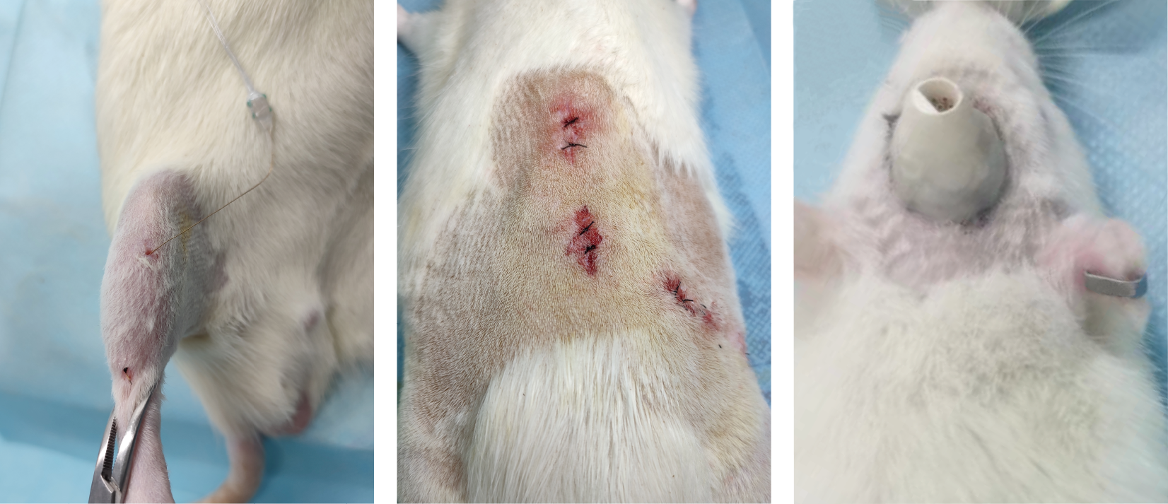
Schematic diagram of minimally invasive implantation of fiber electrodes and photos of skull interfaces.